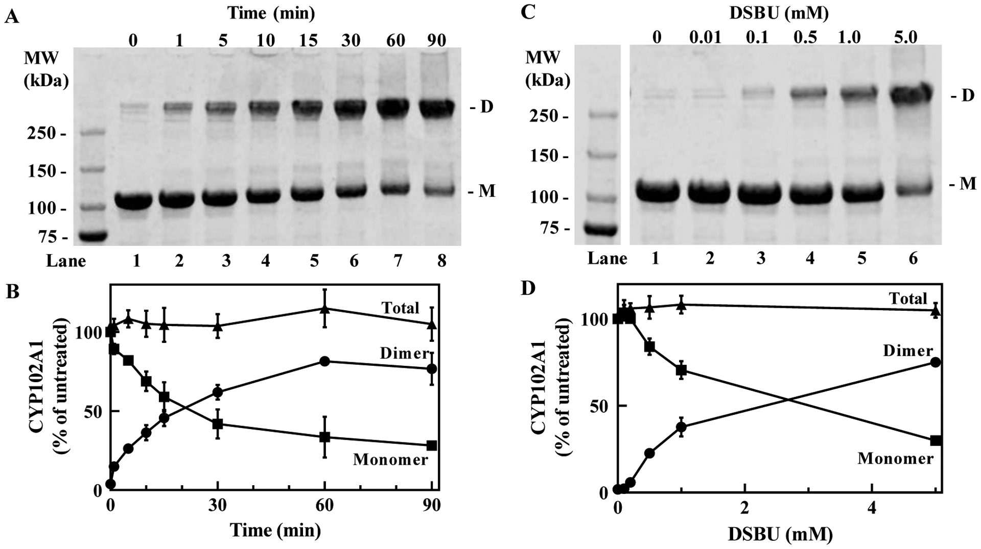
Figure 1. Formation of covalently crosslinked CYP102A1 dimer after treatment with DSBU.

(A), Time-dependent formation of crosslinked CYP102A1 after treatment with DSBU. Full-length CYP102A1 (10 μM) was treated with 0.5 mM DSBU for the indicated duration and aliquots (3 μg) of the reaction mixtures were submitted to SDS-PAGE and stained with Coomassie Blue as described in Methods. D, crosslinked dimeric CYP102A1; M, monomer of CYP102A1. (B), Quantification of bands seen in A. Bands corresponding to the crosslinked dimer (closed circles) and monomer (closed squares) were quantified by densitometric analysis. The sum of the dimer and monomer was also calculated (closed triangle). Mean ± SD derived from three independent reaction mixtures (n=3). (C), Formation of the crosslinked CYP102A1 is dependent on the concentration of DSBU. CYP102A1 was treated with the indicated concentrations of DSBU for 5 min and the reaction mixture analyzed as in A. (D), Quantification of bands seen in C. The amount of dimeric CYP102A1 (closed circles), monomeric CYP102A1 (closed squares), and the sum total (closed triangles) was quantified as in B. Mean ± SD (n=3). Densities determined for all bands are within the linear range of detection.
