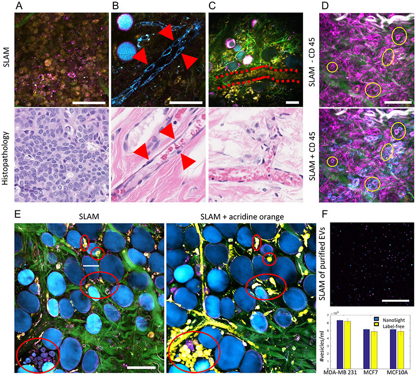
Figure 2. Identification of cells and EVs by comparing label-free (SLAM) images with previously established marker-based methods.
(A) Images of tightly packed, round-shaped, 10-micron-sized tumor cells, (B) aligned and elongated vascular endothelial cells, and (C) stream-like flowing red blood cells in a vessel by SLAM microscopy (upper row) and corresponding H&E-stained histology (lower row). (D) SLAM microscopy of the tumor microenvironment of a living rat without and with CD45 staining for leukocytes. The blue channel was assigned to NADH before staining and assigned to CD45 after staining. (E) Correlation of normal breast cells before and after acridine orange labeling of freshly excised breast tissue. (F) Imaging and characterization of isolated EVs by multiphoton microscopy and NTA. Adapted from previous work using SLAM and full details can be found from the original papers (27,30). Scale bar: 50 μm.

