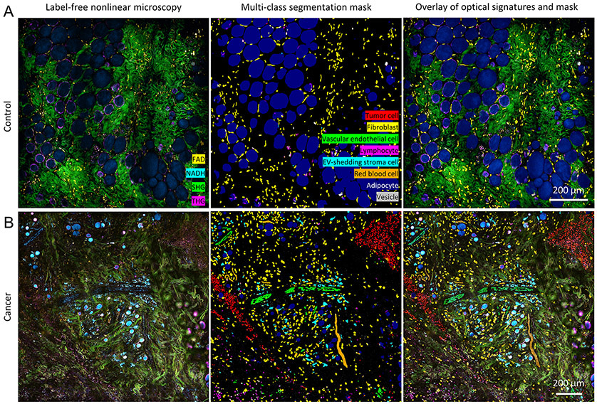
Figure 3. Segmentation results of tumor cells, fibroblasts, vascular endothelial cells, lymphocytes, EV-shedding stroma cells, red blood cells, adipocytes, and EVs from representative label-free images of A) control and B) cancer animals (27).
Scale bar: 200 μm. Red blood cells were labelled and segmented in the form of a blood flow/strip because it was challenging to distinguish them at the single-cell level in the static image, especially for blood flow within the vessels.

