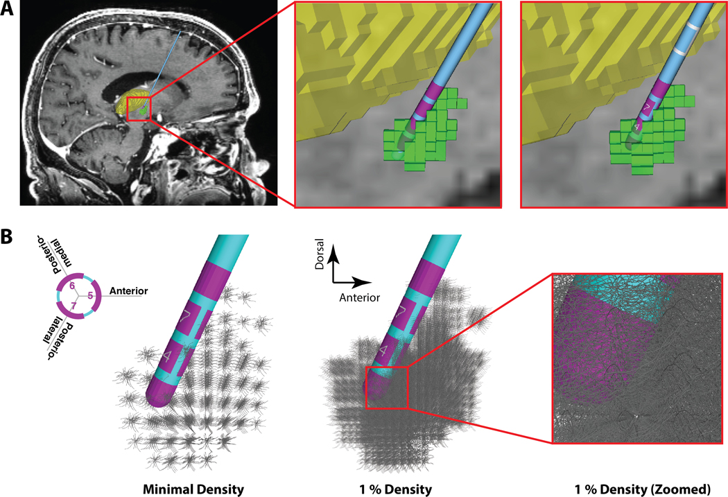
Figure 1.
Anatomical Model. A) Far left panel shows a sagittal view of the patient magnetic resonance image (MRI), deep brain stimulation (DBS) electrode, and 3D anatomical volumes representing the subthalamic nucleus (STN - green) and thalamus (yellow). Middle panel shows a zoomed-in view of the original patient-specific 3389 lead location (pink electrode contacts). Far right panel shows the corresponding 2202 lead location. B) STN neuron models surrounding the DBS electrode. Each grey STN neuron model is displayed with its full 3D geometry of the soma-dendritic architecture. Far left panel, one model neuron is shown in each voxel of the STN volume (185 neurons). Middle panel, 1% density of the STN neurons models (i.e. 2350 out of 235,280 neurons), and right-most panel displays a zoomed-in view around contact 1.

