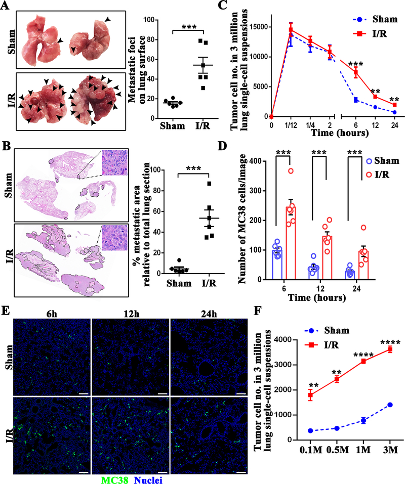
Figure 1.
Surgical stress augments metastatic colonization. (A) Representative images show metastatic pulmonary nodules at day 21 following tumor cell injection in mice subjected to sham or hepatic I/R group. (B) Representative images of H&E staining of lung sections after necropsy in mice subjected to sham or hepatic I/R group. Pathological sections revealed that tumor cells were loosely arranged, with extreme nuclear pleomorphism, hyperchromatic nuclei and nuclear fission. Tumor burden was measured as percent lung tissue replaced by metastatic tumor. (C) Lung tissues were collected at the indicated time after the injection of 106 MC38 cells followed by hepatic I/R or sham surgery. The number of tumor cells in a 3 million lung single-cell suspension was determined by FACS. (D) Quantitation of tumor cells in mouse lungs at 6, 12, and 24 hours after MC38 injection in mice subjected to hepatic I/R compared with the sham group. Points represent the mean ± SEM (n ≥ 9 images). (E) Representative immunofluorescence images by confocal microscopy of mice lung sections showing tumor cells at the times indicated following the injection of MC38 cells. Green: MC38; Blue: Nuclei. Scale bar, 100μm. (F) Lung tissues were collected at 12 hours after the injection of different dose of MC38 cells (105, 5 × 10⁵, 106 and 3 × 10⁶) followed by hepatic I/R or sham surgery. The number of tumor cells in a 3 million lung single-cell suspension was determined by FACS. Data are presented as mean ± SEM from n = 3–6 mice per group. **P<0.01, ***P<0.001 and ****P<0.0001.

