Figure 2.
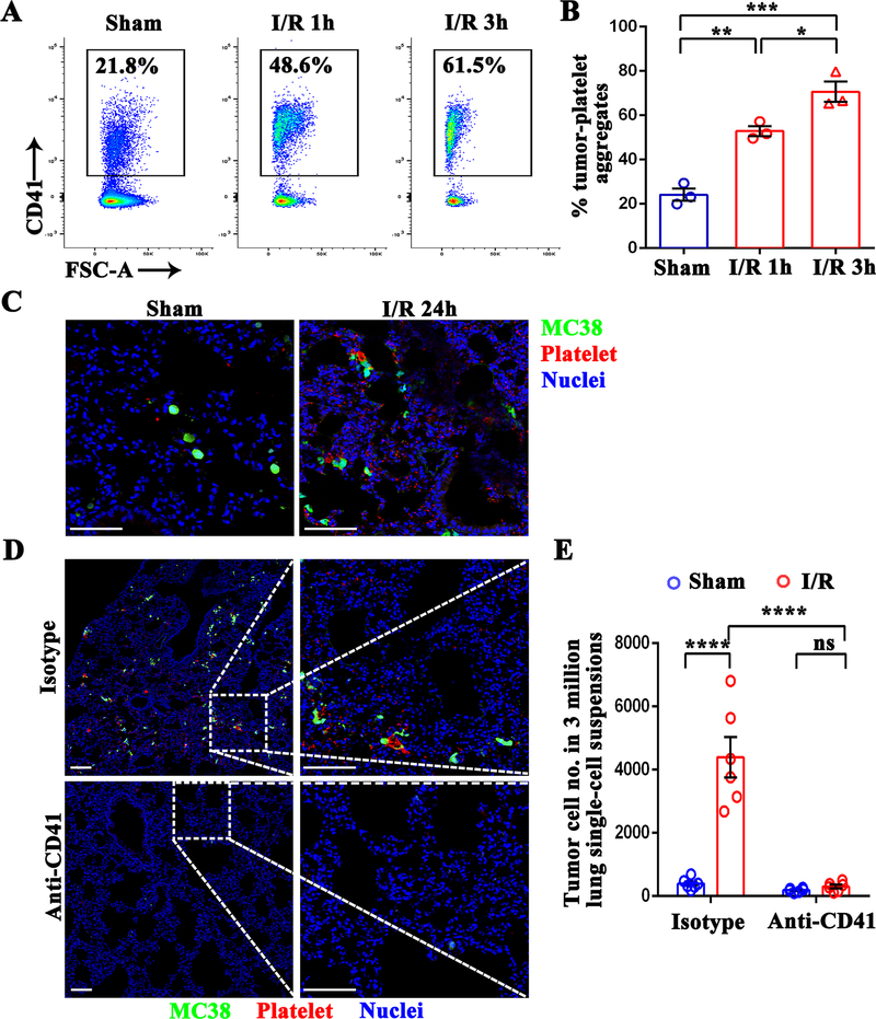
Hepatic I/R promotes the formation of platelet-tumor cell aggregates and subsequent distant metastasis. (A) Representative flow cytometry showing platelet-tumor cell aggregates. Following 1.5 hours of hepatic ischemia and 1 or 3 hours of reperfusion, platelets were extracted from I/R or sham mice, and then co-cultured with CFSE-labeled MC38 for 10 minutes. (B) From the flow cytometry results in A, platelet-MC38 aggregates were calculated as a percentage of total cancer cells. (C) Representative immunofluorescence images showing colocalization pattern between tumor cells and platelets at 24 hours post-injection. Platelets were stained with anti-CD41-PE. Green: MC38; Red: platelet; Blue: Nuclei. Scale bar, 100μm. (D) WT mice were injected intraperitoneally with either platelet-depleting anti-CD41 antibody or IgG isotype followed by tumor cell injection and hepatic I/R. Representative immunofluorescence images by confocal microscopy of mice lung sections showing tumor cells arrested at 24 hours post-injection. Green: MC-38; Red: Platelet; Blue: Nuclei. Scale bar, 100μm. (E) FACS quantitative data of MC38 cell numbers in the lungs of mice at 12 hours post-injection. Data are presented as mean ± SEM from n = 3–6 mice per group. ns: not significant, *P<0.05, **P<0.01, ***P<0.001 and ****P<0.0001.

