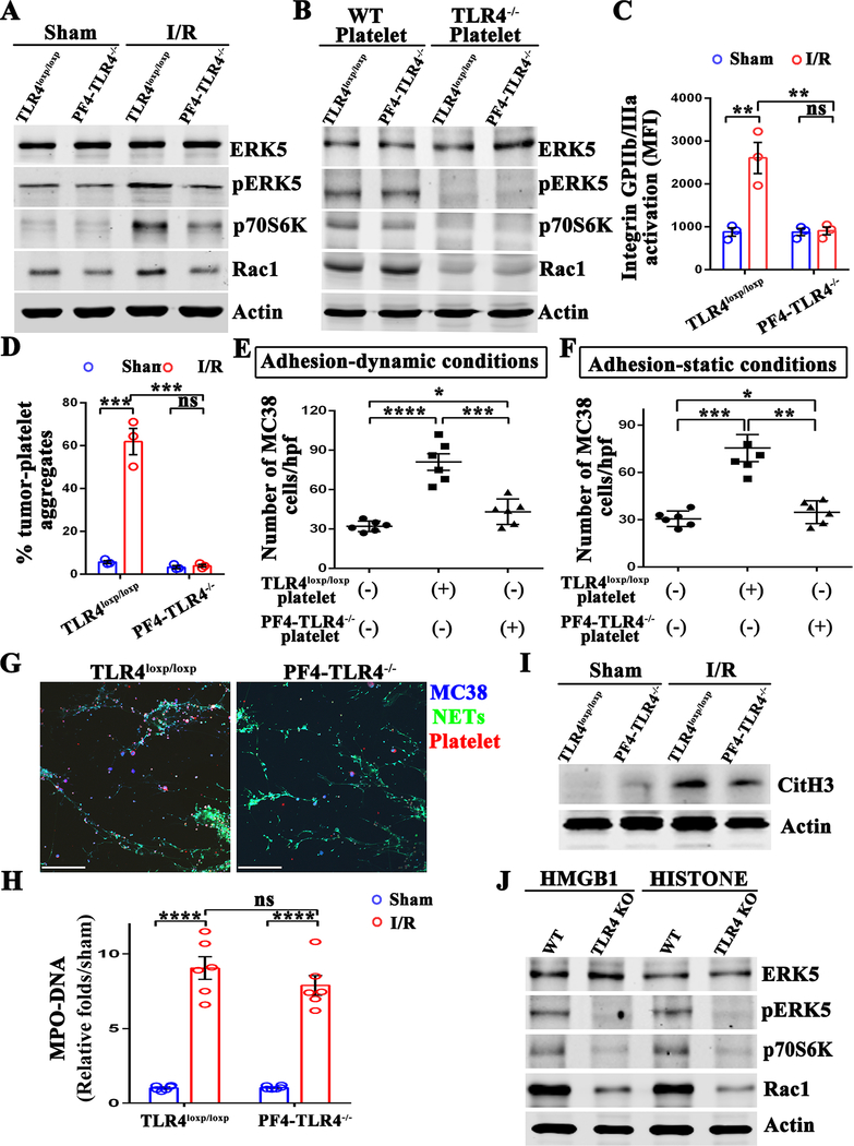
Figure 6.
Platelet TLR4 is essential for activation of ERK4 and NET-mediated capturing of tumor cells. (A) Western blot analysis of indicated proteins in the platelets extracted from TLR4 flox (TLR4loxp/loxp) and platelet-specific TLR4-KO (PF4-TLR4−/−) mice after either sham laparotomy or 1.5 hours of hepatic ischemia and 3 hours of reperfusion. (B) Platelets obtained from wild type and TLR4−/− mice were adoptively transferred into platelets-depleted TLR4loxp/loxp and PF4-TLR4−/− mice. Western blot analysis of indicated proteins in the platelets extracted from the above mice after 1.5 hours of hepatic ischemia and 3 hours of reperfusion. (C) Flow cytometry analysis of integrin activation in platelets extracted from TLR4loxp/loxp and PF4-TLR4−/− mice after either sham laparotomy or hepatic I/R. (D) Aggregation capacity of platelets from C with MC38. (E and F) Quantitation of tumor cells that adhered to NETs in the presence of TLR4loxp/loxp or PF4-TLR4−/− platelets under static and dynamic conditions. (G) Representative immunofluorescent staining of tumor adhesion to NETs under static conditions. Blue: MC-38; Green: NETs; Red: Platelet. Scale bar, 100μm. (H) Serum MPO-DNA complex levels were assessed in TLR4loxp/loxp and PF4-TLR4−/− mice after either sham laparotomy or 1.5 hours of hepatic ischemia and 6 hours of reperfusion. (I) Western blot analysis of Cit-H3 protein level in mice from H. (J) Platelets isolated from wild type and TLR4−/− mice were stimulated with recombinant HMGB1 (5ug/ml) or histone protein (25ug/ml) for 6 hours. Western blot analysis of indicated proteins in these platelets. Data are presented as mean ± SEM from n = 3–6 mice per group. ns: not significant, *P<0.05, **P<0.01, ***P<0.001 and ****P<0.0001.

