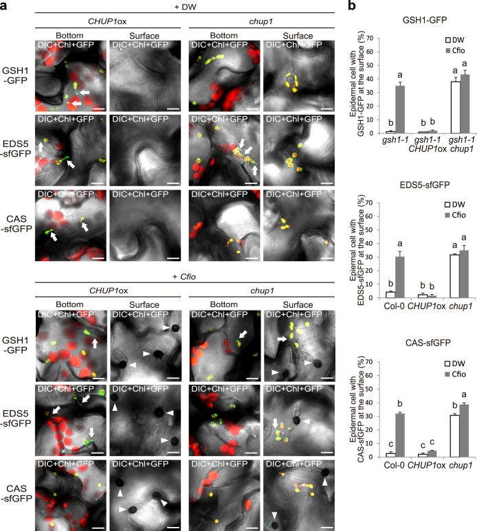
Fig. 6. Localization pattern of GSH1, EDS5, and CAS tightly linked to epidermal chloroplasts and the ECR.
a Localization pattern of GSH1, EDS5, and CAS proteins in CHUP1ox and chup1 plants. The transgenic gsh1 CHUP1ox and gsh1 chup1 plants expressing GSH1-GFP under its own promoter, and CHUP1ox and chup1 plants expressing EDS5-sfGFP and CAS-sfGFP under the CaMV 35S promoter were incubated with Cfio and observed at 2 dpi. DW was used as a control. The chloroplasts were visualized based on chlorophyll autofluorescence. The white arrowheads and arrows indicate melanized appressoria and stromules, respectively. Scale bar, 10 µm. b Quantification of epidermal cells with surface fluorescence of GFP/sfGFP-labeled immune components. Cfio-inoculated cotyledons of the indicated Arabidopsis were analyzed at 2 dpi. A total of 100 cells in contact with the melanized appressorium were observed. As a control, DW was spotted. The means and SE were calculated from three independent plants. Means not sharing the same letter are significantly different (P < 0.05, two-way ANOVA with Tukey’s HSD test).

