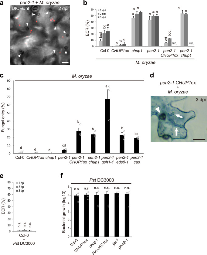
Fig. 9. The ECR contributes to preinvasive NHR against M. oryzae.
a ECR after inoculation of M. oryzae. M. oryzae was inoculated onto cotyledons of pen2-1 plants, and the ECR was investigated at 2 dpi. The chloroplasts were visualized based on chlorophyll autofluorescence. b Quantification of ECR against M. oryzae. ECR was investigated at 1, 2, and 3 dpi. A total of 100 cells in contact with the melanized appressorium were observed. N.D.: not determined due to damages of epidermal cell by fungal invasion. c Entry rate of M. oryzae into epidermis at 3 dpi. A total of 100 melanized appressoria were investigated. d Epidermal cell death caused by appressorium-mediated entry of M. oryzae at 3 dpi. The inoculated plants were subjected to TB staining and observed microscopically using x40 objective lens. The arrowheads and arrows indicate melanized appressoria and invasive hyphae, respectively. Scale bar, 20 µm. The means and SE were calculated from three independent plants. Means not sharing the same letter are significantly different (P < 0.05, one-way (c) or two-way (b) ANOVA with Tukey’s HSD test). e Quantification of ECR against Pseudomonas syringae pv. tomato (Pst) DC3000. Cotyledons of Col-0 were drop-inoculated with Pst DC3000, and ECR was investigated at 1, 2, and 3 dpi. A total of 100 epidermal pavement cells were observed. The means and SE were calculated from three independent plants. f Growth of Pst DC3000 in Arabidopsis cotyledons. Cotyledons of indicated Arabidopsis were drop-inoculated with Pst DC3000, and incubated for 4 days. The number of bacteria in eight cotyledons obtained from four independent plants was plotted on a log10 scale. The means and SE were calculated from three independent experiments. n.s. not significant (*P < 0.05, one-way (f) or two-way (e) ANOVA with Tukey’s HSD test).

