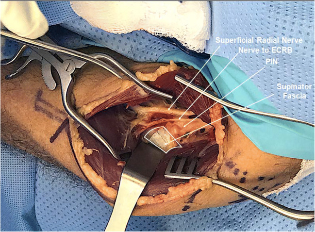
Fig. 9.
This image demonstrates the neural structures between the brachioradialis anteriorly and ECRL posterolaterally. The wide white band deep to the ECRL is the supinator fascia and posterior interosseus nerve (PIN) can be seen diving deep to the supinator fascia. Just anterior to the PIN a branch can be seen which is the nerve to extensor carpi radialis brevis (ECRB). Anterior to this, deep to the brachioradialis is the superficial radial nerve that has branched with the PIN from the radial nerve proximal to this exposure. ) (Image Courtesy of Louis Catalano, MD)

