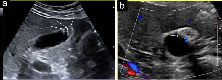
Fig. 9.
a The sonographic images show the localised type of GA as a focal thickening localised in the fundus. The thickening projects into the lumen and appears hypoanechoic with hyperechoic cholesterol crystals generating comet-tail reverberation artifacts (white arrows). b The sonographic images show the localised type of GA as a focal thickening localised in the fundus. The thickening projects into the lumen and appears hypoanechoic with hyperechoic cholesterol crystals generating twinkling artifacts on the colour Doppler exam (yellow arrow)

