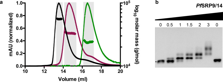
Fig. 2. Assembly of the PfAlu domain.
a Profiles of size-exclusion chromatography coupled to multi-angle light scattering of PfAlu118 RNA (magenta), PfSRP9/14 (green), and the complete PfAlu domain comprising PfAlu118 RNA and PfSRP9/14 (black) are shown. Molar mass distribution across the respective peaks is marked and fractions used for obtaining an average molar mass are highlighted in gray. b EMSA showing quantitative shifts of the PfAlu118 RNA upon titration with increasing amounts of PfSRP9/14. The asterisk (*) indicates unbound RNA. The molar-fold excess of PfSRP9/14 over PfAlu118 RNA is indicated on the top. The uncropped gel image is included in Supplementary Data 1.

