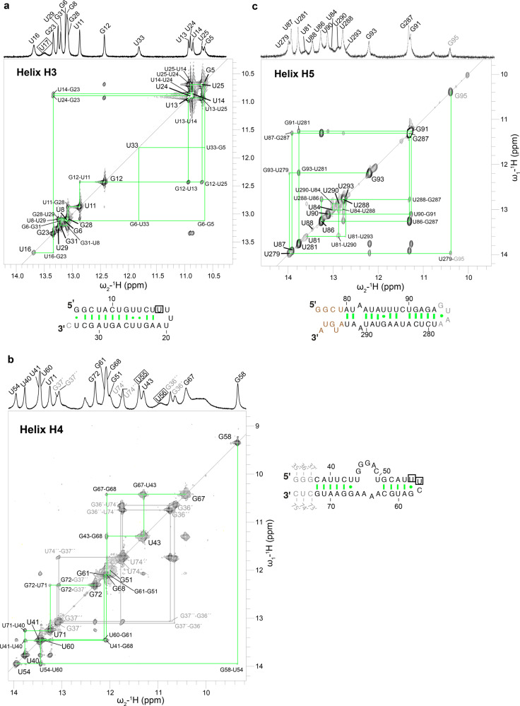
Fig. 3. PfAlu RNA secondary structure.
Imino regions of 1D and 2D 1H,1H-NOESY spectra of helices H3, H4, and H5 are shown in a–c, respectively. RNA sequences of the corresponding helices are also shown. Nucleotides colored in gray have been added artificially to the Alu RNA while those in brown could not be assigned. Wobble base pairs are connected by a dot while canonical Watson–Crick base pairs are connected by a hyphen. Resonances, which did not give imino cross-peaks to neighboring bases and were assigned by exclusion, are boxed. Helix H4 resonances exhibiting alternative conformations are numbered in gray.

