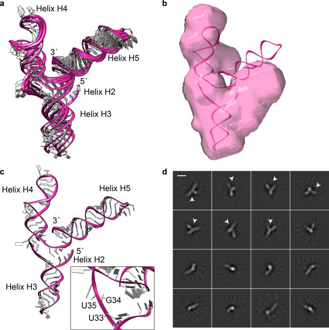
Fig. 5. Structure analyses of the PfAlu118 RNA.
a NMR-SAXS-based atomic models of PfAlu118 RNA. b Ab inito model of PfAlu118 RNA superimposed with the top scored PfAlu118 RNA NMR-SAXS model calculated by SREFLEX. c Top scored PfAlu118 RNA NMR-SAXS model with inset showing the UGU motif exposed at the three-helix junction. d Single-particle cryo-EM analysis of PfAlu118 RNA shown with representative reference-free 2D-class averages. Arrows mark class averages where the Y-shaped architecture of the RNA is visible. Scale bar represents 30 Å.

