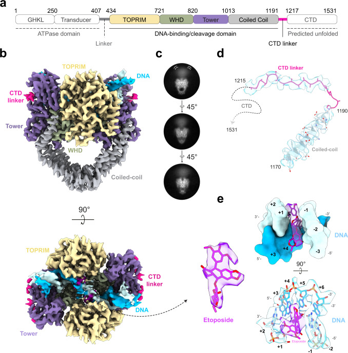
Fig. 1. Cryo-EM structure of the hTopo IIα etoposide-poisoned DNA-binding/cleavage domain.
a Schematic representation of the hTopo IIα DNA-binding/cleavage domain. b Cryo-EM structure of the DNA-binding/cleavage domain in State 1 solved at 3.6 Å resolution. The structure is colored as in a, the DNA is colored in blue. c 2D classes of hTopo IIα in different orientations showing the flexibility of the ATPase domain. d EM density around the last coiled-coil helix and the CTD linker. e Zoom on the EM density of the etoposide and its binding site intercalating DNA bases +1 and −1.

