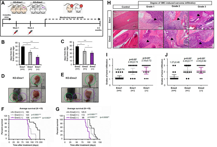
Fig. 5. Model of sarcomagenesis in vivo induced by 3MC in the different cohorts of Emx1 or Emx2 KO mice.
A Scheme of the experimental procedure and monitoring of intramuscular carcinogenic induction mediated by 3MC in murine models of Emx1 and Emx2. Each cohort comprised 15 mice. B, C Graph of the days since the first tumor sign in each cohort of Emx1 (B) and Emx2 (C). F, G Survival curve with respect to time post treatment with 3MC in Emx1 (F) and Emx2 (G). D, E Representative photographs of the control and tumor limbs of each Emx1 (D) and Emx2 (E) cohort. The p-value of the survival graphs was obtained using the LogRank test. The rest of the analysis was performed with Student’s t-test (*p < 0.05; **p < 0.01; ***p < 0.001). H–J Effect of Emx1 or Emx2 depletion on the tumor invasiveness of the bone marrow of the femur in the 3MC-induced sarcomas. In H, representative micrographs of the three grades into which the evolution of the tumor infiltration of sarcoma induced by 3MC in the bone marrow has been divided with respect to the control tissue. The points of initiation or progression of the infiltration are highlighted with the arrowhead. The bar indicates 200 µm. I, J Graphs of the degree of tumor infiltration measured in the Emx1 (I) and Emx2 (J) KO line cohorts. Each cohort had 15 mice. Statistical analysis was performed with Student’s t-test (*p < 0.05; **p < 0.01; ***p < 0.001).

