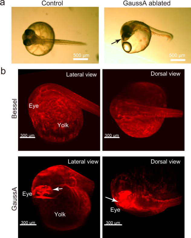
Fig. 6. Phototoxicity assessment.

a Bright field view of the 3 dpf embryos of the β-actin-DsRed2 strain after the phototoxicity experiment. Control indicates a gel-embedded embryo, which does not show significant damage. For the ablation experiment, the time-course 4D imaging was performed in 5-min intervals with 200 ms exposure/slice with 2 μm steps, 2048 × 2048 pixels, ~600 slices. The arrow shows the air bubble created around the eye. b 3D view of the embryos viewed from the lateral and dorsal side. White arrows indicate strong fluorescence emitted from the retinal pigments. a, b n = 3 biologically independent β-actin-DsRed2 strain embryos are examined for the different beam conditions of phototoxicity experiment.
