Figure 8.
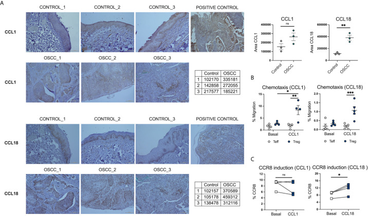
CCL18 is augmented in histological samples of OSCC patients and also induce CCR8 expression in Teff. (A) Representative histological staining of CCL1 and CCL18 in a biopsy from a patient with OSCC and a patient without malignancy, using colon carcinoma as a positive control for CCL1 and melanoma as a positive control for CCL18. (B) Semi-quantification of area for CCL1 and CCL18 staining by ImageJ, data is presented as mean ± SEM using individual values described in the tables (Unpaired t test). (C) Percentage of migrated memory Teffs and Tregs to recombinant chemokines CCL1 and CCL18. Sorted Cell trace violet+ Memory Teffs (5×10⁴) and unstained memory Tregs (5×10⁴) were placed in the top chamber of a 5-μm-pore Transwell filter system. Bottom chambers were filled with media only, CCL1 or CCL18, (all 0.5 ug/mL). The percentage of migration for each subset was calculated as (number of cells in the bottom chamber after 1 h × 100)/initial number of cells in the top chamber. Data are presented as mean ± SEM using scatter dot plots (Paired t test). (C) CCR8 expression was measured in pre-activated memory Teffs (1x10⁵) cultured with media only, or media with CCL1 or CCL18, (all 0.5 ug/mL) for 72h, data are presented using individual symbols with paired lines (Paired t test). For all statistical tests, ∗∗∗p < 0.001, ∗∗p < 0.01 and ∗p < 0.05 were considered significant. ns, not significant.

