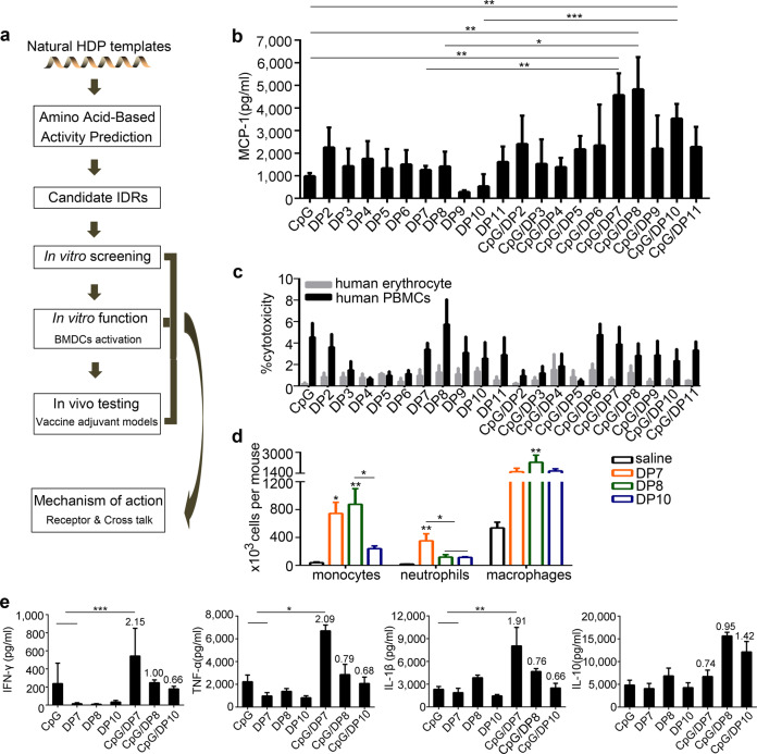
Fig. 1. In vitro preliminary screening of defense peptide candidates.
a A representative flowchart of the IDR design. b PBMCs were stimulated with CpG (20 µg/ml), DP2-11 (40 µg/ml), or CpG: DP2-11 (1:2; wt/wt) formulations for 24 h. The secretion of MCP-1 was detected by ELISA. c PBMCs or human erythrocytes were incubated with CpG (20 µg/ml), DP2-11 (40 µg/ml), or CpG/DP2-11 (1:2; wt/wt) formulations for 24 h. After that, the supernatants were collected, and LDH release from PBMCs and total hemoglobin release from red blood cells were measured. d DP7, DP8, and DP10 induced the significant recruitment of neutrophils (Gr1+F4/80−), monocytes (F4/80+Gr1+), and macrophages (F4/80+CD11b+) to the injection site at 24 h post-injection (N = 5/group). e PBMCs were stimulated with CpG (20 µg/ml), DP7 (40 µg/ml), DP8 (40 µg/ml), DP10 (40 µg/ml), CpG/DP7, CpG/DP8 or CpG/DP10 (1:2; wt/wt) formulations for 48 h. The secretion of the indicated cytokines was detected. N = 3 per group. Bars represent means and SEM. The numbers in the graph indicate the synergistic values. *P < 0.05; **P < 0.01; ***P < 0.001.

