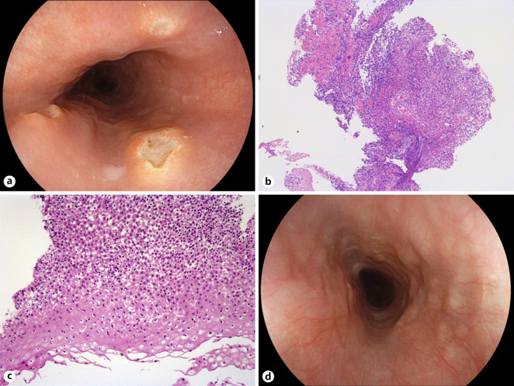
Fig. 1.
a Esophagogastroduodenoscopy (EGD) demonstrating esophageal ulcers with an estimated maximum size of 8 mm and slightly raised borders. Note that all esophageal mucosal injury lesions are located at the same height within the esophagus at 27 cm from the incisors (most likely representing physiological narrowing due to the left main bronchus). b, c Biopsies from the ulcer bed and rim indicate bland ulceration with a dense inflammatory infiltrate and epithelial edema (H&E. ×5, ×10, respectively). In addition, there is no evidence for viral esophagitis as substantiated by negative herpes simplex virus type 1 and cytomegalovirus immunohistochemistry (not shown). d Complete ulcer healing on repeat EGD 14 days later.

