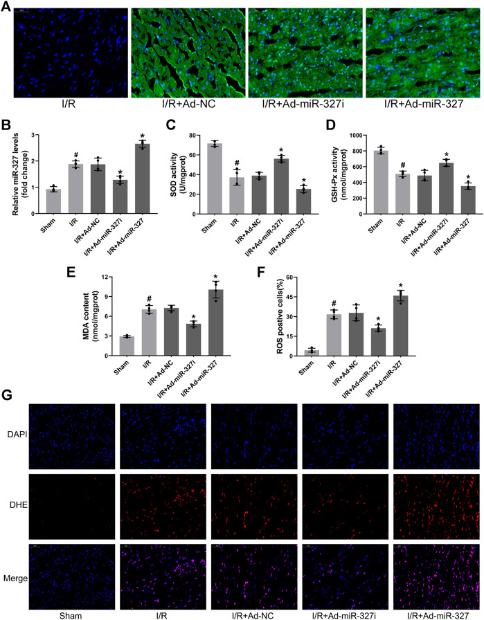
FIGURE 8.
Effects of miR-327 on oxidative stress in MI/RI rats. Representative images of immunofluorescence microscopy after transfection of recombinant adenovirus or NS injection, DAPI-labeled nuclei of cardiomyocytes (blue), EGFP(green) and merged (A). Relative expression of miR-327 in MI/RI rats (B). The levels of SOD (C), GSH-Px (D), and MDA (E) in heart tissues of MI/RI rats. DHE staining for detecting intracellular ROS generation in heart tissues of MI/RI rats (G) in each group, and the analysis of ROS positive cells by Image-Pro Plus (F). Data are expressed as the mean ± SD (n = 4); #p < 0.05 versus the Sham group; *p < 0.05 versus the Ad-NC group.

