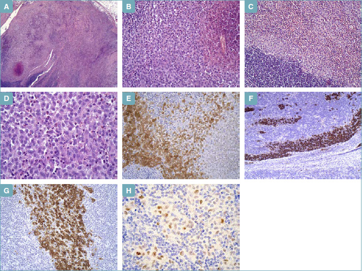
Figure 2.

Lumpectomy. (A) Lymph node with pale areas, with central necrosis (hematoxylin-eosin). (B) Medium sized round-oval cells, with grooved, folded, indented nuclei, fine chromatin, small nucleoli, delicate nuclear membrane, and abundant pale eosinophilic cytoplasm, in aggregates with central necrosis (hematoxylin-eosin). (C) Langerhans cells mixed with a large number of eosinophilic granulocytes (hematoxylin-eosin). (D) Langerhans cells at higher magnification (hematoxylin-eosin). (E) S100 protein (immunohistochemistry) positive in Langerhans cells. (F) CD1a (immunohistochemistry) positive in Langerhans cells (intra-sinusoidal distribution). (G) Langerin/CD207 (immunohistochemistry) positive in Langerhans cells. (H) Cyclin D1 (immunohistochemistry) positive in Langerhans cells.
