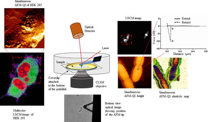
Figure 1.
Schematic showing the multiplexed data arising from correlative atomic force-confocal microscopy. AFM in quantitative imaging (QI™) mode quantifies elasticity, adhesion (extracted from nanoscopy curves, top right) and surface topography, while confocal can localize multiple fluorescently labeled molecules within the sample (bottom left). Simultaneous imaging can be conducted in live, physiologically relevant conditions in real-time. This method can be easily extrapolated to characterizing human-pathogen interactions, in the presence or absence of therapeutics, at the single-cell level. This figure has been reproduced from Bhat et al. (2018b) with permission from Scientific Reports.

