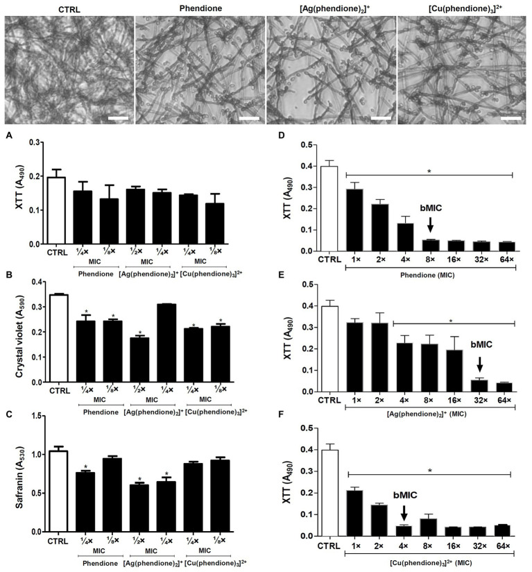
Figure 1.
Effects of phendione and its metal complexes on P. verrucosa biofilm formation and maturation. Conidia (1 × 10⁶) were added to 96-well plates containing Roswell Park Memorial Institute (RPMI) medium supplemented with sub-MIC concentrations of each test compound. After incubation for 72 h at 37°C, (A) cell viability, (B) quantification of biomass, and (C) extracellular matrix were evaluated, as described in Material and Methods. The same conidia density (D–F) was placed to interact with polystyrene for 72 h in RPMI medium. Then, concentrations varying from MIC to 64 × MIC of each test compound were added and the plates incubated for additional 48 h. The MIC90 values of the biofilm (bMIC90) were detected using the XTT reduction assay. Systems containing non-treated conidia were also prepared (CTRL, control). The eluent (dimethyl sulfoxide, DMSO) did not affect fungal growth (data not shown). The symbol (*) highlights the MIC values that caused a statistically significant reduction on each evaluated parameter in relation to the respective control (p < 0.05). Representative images of biomass (crystal violet-stained) formed by P. verrucosa non-treated (CTRL, control) and treated with phendione (¼ × MIC) and its silver(I) (½ × MIC) and copper(II) (¼ × MIC) complexes. Bar: 20 µm.

