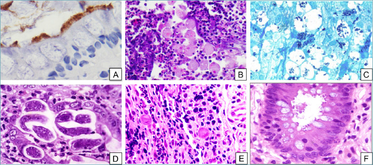
Figure 1.

Infectious colitis. (A) Intestinal spirochetosis. The fuzzy luminal border is highlighted by immunohistochemistry for Treponema pallidum (peroxidase-diaminobenzidine, 40x). (B) Entamoeba histolytica trophozoites containing engulfed erythrocytes can be seen among neutrophils and inflammatory debris (H&E, 20x). (C) Grocott’s stain makes Histoplasma capsulatum stand out as intensely-stained small oval structures within the cytoplasm of macrophages (Grocott’s methenamine silver stain, 20x). (D) Strongyloides stercoralis can be found lurking in the crypts (H&E, 20x). (E) CMV-infected cells stand out as markedly enlarged cells with typical “owl’s eye” intranuclear inclusions (H&E, 20x). (F) Cryptosporidium parvum forms small round bodies on the luminal border of enterocytes (H&E, 20x). !
