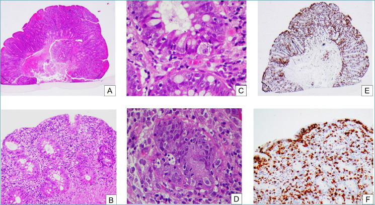
Figure 4.

Autoimmune enteropathy. (A) At low power the mucosa appears reactive, with regenerative crypts and an hypercellular lamina propria (H&E, 2x). (B) Crypts are regenerative, with mucin depletion and surface epithelial damage (H&E, 10x). (C) On closer inspection, there are prominent apoptotic bodies in the surface and crypt epithelium (H&E, 40x). (D) Sometimes, satellite lymphocytes can be seen around the apoptotic bodies (H&E, 40x). (E) CD3 immunohistochemistry reveals a greatly increased T lymphocyte population (H&E, 2x). (F) The T lymphocytes can be seen in the lamina propria as well as in crypt and surface epithelium (H&E, 20x).
