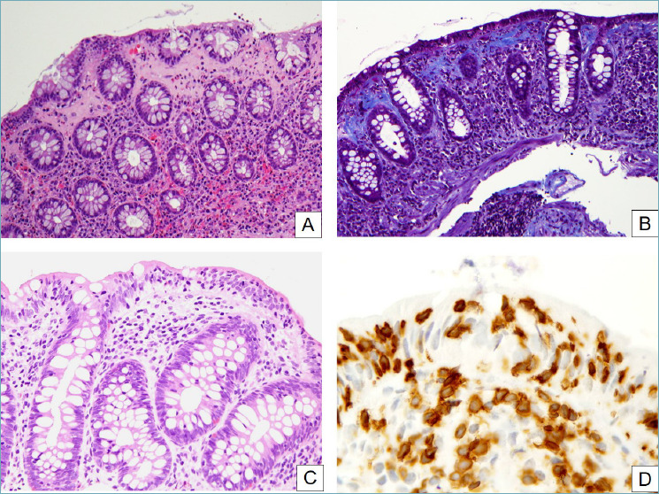
Figure 5.

Microscopic colitis. (A) In collagenous colitis, a thick and irregular collagen band underlies the surface epithelium and often entraps inflammatory cells. Surface epithelial detachment, shown here, is also typical. The underlying lamina propria is inflamed and shows an increased amount of eosinophils (H&E, 20x). (B) A Masson trichrome clearly highlights the irregularly thickened subepithelial collagen band (Masson trichrome, 20x). (C) In lymphocytic colitis, numerous T lymphocytes infiltrate the gland epithelium (H&E, 20x). (D) CD3 stain clearly highlights the pathologically increased intraepithelial lymphocytes (peroxidase-diaminobenzidine, 40x).
