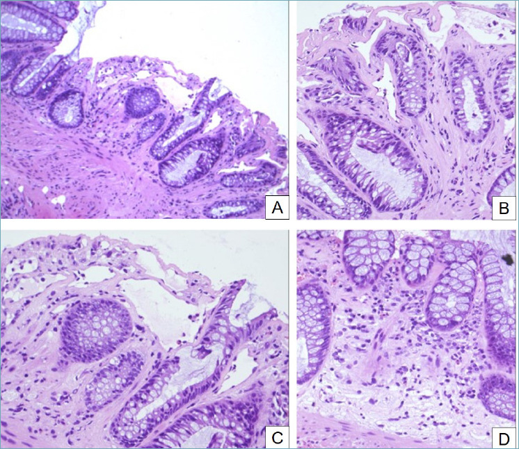
Figure 6.

Radiation-induced colitis. (A) Crypt distortion, dilation and atrophy are evident (H&E, 10x). (B) Fibrosis of lamina propria, hyperplasia of crypts and dilated vascular channels (H&E, 20x). (C) Crypt atrophy, apoptotic bodies, goblet cell depletion and ectasia of small vessels are typical of radiation-induced colitis (H&E, 20x). (D) The lamina propria shows an inflammatory infiltrate that tends to decrease in long-standing cases (H&E, 20x).
