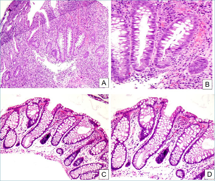
Figure 7.

Segmental colitis associated with diverticular disease. (A) In this case, crypt distortion, cryptitis and basal plasmacytosis dominate the picture (H&E, 10x). (B) Detail of the previous image. Active inflammation and cryptitis can be appreciated, as well as the increased number of plasma cells and eosinophils in the inflammatory infiltrate (H&E, 40x). (C, D) In this other case, inflammation is much less florid, while crypt distortion and stromal edema are predominant (H&E, 20x and 40x respectively).
