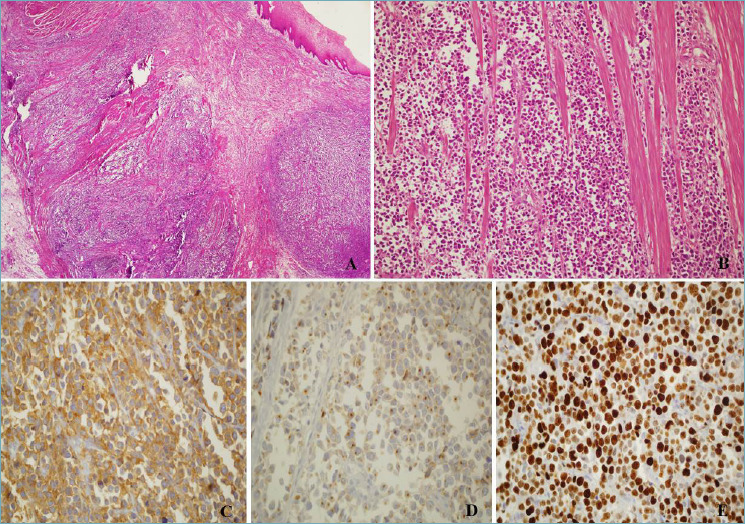
Figure 1.

(A) Esophageal small cell neuroendocrine carcinoma undermining normal squamous esophageal mucosa; H&E, magnification 4x. (B) Small cell neuroendocrine carcinoma infiltrating the muscular wall of the esophagus; H&E, magnification 10x. (C) Diffuse positivity of neoplastic cells for synaptophysin, magnification 20x. (D) Faint, dot-like, positivity for Chromogranin A; magnification 20x. (E) High proliferative index, 80% (Ki-67 stain), magnification 20x.
