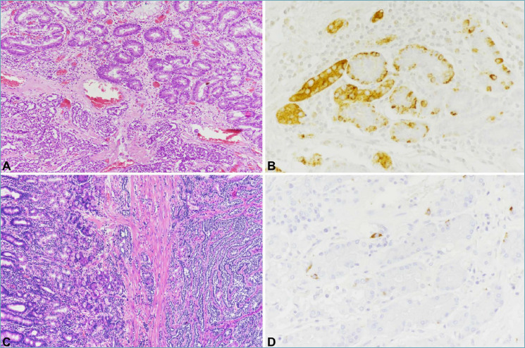
Figure 2.

Type 1 ECL-cell NET is composed of well-differentiated cells arranged in small microlobular and/or trabecular structures (A, bottom). The peritumoral oxyntic mucosa (A, top) is atrophic with diffuse intestinal metaplasia and shows ECL-cell linear and micronodular hyperplasia, easily detected with chromogranin A immunostaining (B). Type 3 NETs is composed of well differentiated neuroendocrine cells as well, deeply invading the gastric wall (C, right). Peritumoral oxyntic mucosa is normal (C, left) without ECL-cell proliferations (D).
