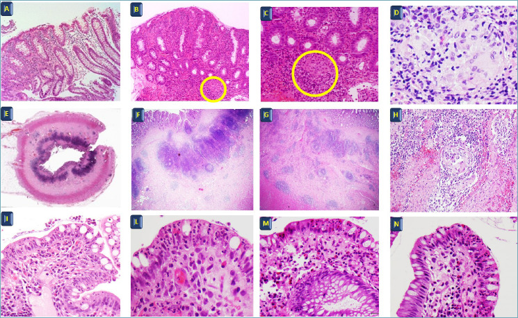
Figure 2.

Crohn’s disease. (A) Patchy inflammation, (B-C) granuloma, (D) granuloma. H&E A-B: 20x, C: 40x, D: 60x. (E, F, G, H) Crohn’s disease surgical specimen, inflammation penetrating the different layers of the terminal ileum H&E E: 4x, F: 10x, G: 20x, H: 40x. (I, L, M, N) Early Crohn’s disease, focal superficial inflammation in the terminal ileum. H&E I: 10x, L: 40x, M: 20x; N 40x.
