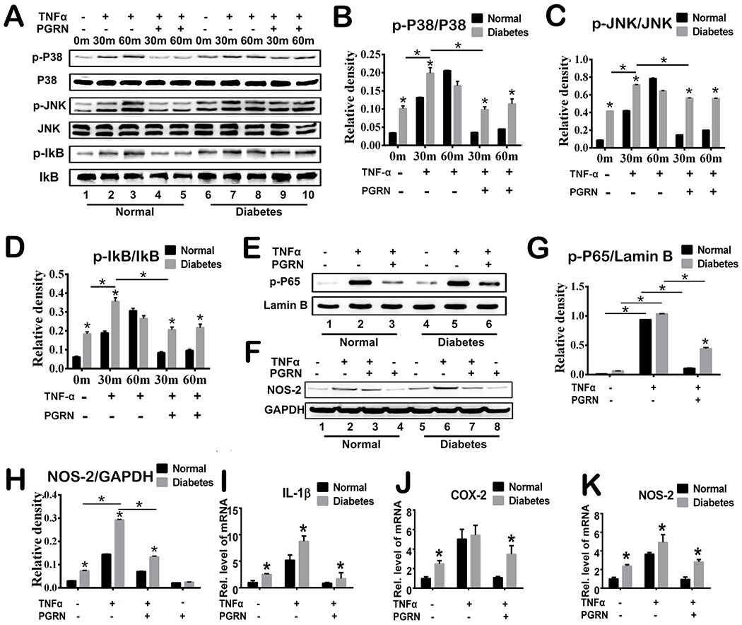
Fig.5. PGRN promotes diabetic fracture healing by inhibiting TNFα-mediated catabolic responses.

(A) Primary mouse bone marrow cells were incubated with or without TNFα (10ng/ml) in the presence or absence of PGRN (200 ng/ml) for 48 hours, and phosphorylation and expression of the indicated signaling molecules were determined by Western Blotting. (B-D) Relative band density of p-P38/P38, p-JNK/JNK and p-IkB/IkB based on Western Blotting. (E) Phosphorylation and expression of p-65 was determined by Western Blotting assay. Lamin B is employed as loading control. (F) Expression of NOS-2 and GAPDH (serving as a loading control) was determined by Western Blotting assay. (G-H) Relative band density of p-P65/lamin B and NOS-2 based on Western Blotting. (I-K) Primary mouse bone marrow cells were cultured without or with TNFα in absence or presence of PGRN for 6 hours. Transcriptional levels of IL-1β, COX-2 and NOS-2 were determined by Real-time PCR assay. Two-way ANOVA was used for the statistical analysis. The values are the mean ±SD. *p < 0.05 vs. control group.
