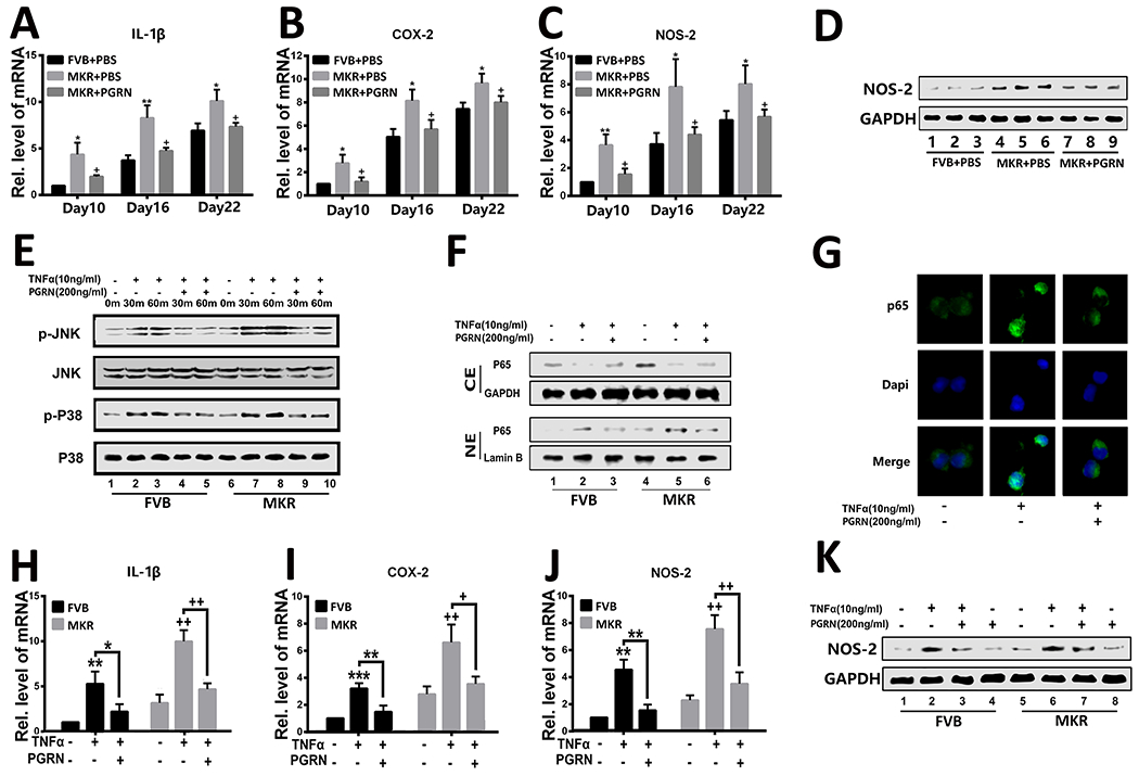
Figure.5. PGRN suppresses TNFα-induced inflammation in T2DM.

(A–C) Transcriptional levels of IL1B, COX2, and NOS2 in the callus of Bonnarens and Einhorn fracture mice on day 10 post-surgery as determined by reverse transcriptase-polymerase chain reaction (RT-PCR). (D) Protein level of NOS-2 in the callus of Bonnarens and Einhorn fracture mice on day 10 post-surgery, as assessed by western blotting. (E) Activation of P38 (p-P38) and JNK (p-JNK) in primary bone marrow cells cultured without or with TNFα and/or PGRN for various time points was determined by western blotting. Total P38 and JNK served as internal controls. (F) P65 was determined by western blotting assay in primary bone marrow cells that were cultured with TNFα, PGRN, or both. (G) Immunofluorescence staining of P65 in primary bone marrow cells of MKR mice in the presence of TNFα or PGRN plus TNFα. (H–J) Expression of IL1B, COX2, and NOS2 in primary bone marrow cells of FVB and MKR mice, determined by RT-PCR. (K) western blotting assay of NOS-2 in primary bone marrow cells cultured in the presence of TNFα, PGRN, or both for 48 hours. The values shown represent the mean ± standard deviation. *p < 0.05 and ** p < 0.01 vs. control group.
