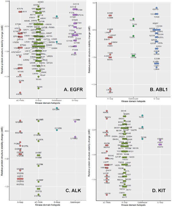
Figure 4.

The relative protein structure stability change by a KD mutation in four TKs. These plots are drawn for the mutations that decreased the relative protein structure stability of EGFR (A), ABL1 (B), ALK (C) and KIT (D). Note there is no αC-helix for ABL1.
