Figure 3.
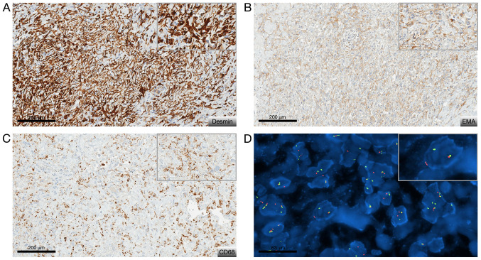
Immunohistochemical staining and FISH analysis results. Characteristic (A to C) phenotypical and (D) molecular features of angiomatoid fibrous histiocytoma. (A) Immunohistochemical view revealing diffusely positive staining for desmin (D33 clone; magnification, x20), (B) patchy staining for EMA (E29 clone; magnification, x20), and (C) CD68 (KP1 clone; magnification, x20). (D) FISH view revealing EWSR1 rearrangement. Original magnification, x63. The top right corner frame shows a 1.5X higher magnification. EMA, epithelial membrane antigen; FISH, fluorescence in situ hybridization.

