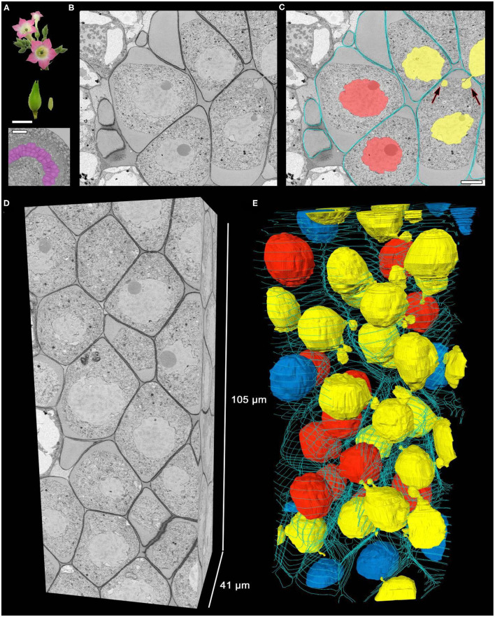
Figure 1.
Three-dimensional reconstruction of male meiocytes in a tobacco anther. (A) Tobacco flowers, a flower bud, an anther, and transverse section of an anther locule. The purple color indicates meiocytes. (B,C) A meiocyte micrograph obtained by SBF-SEM at pachytene. An original image (B) and the same image after cell walls and nuclei were labeled (C). (D,E) Three-dimensional reconstructions of a scanned tissue fragment from 2,626 serial images. The whole unlabeled tissue fragment (D) and labeled nuclei and cell walls (E). Yellow denotes migrating nuclei, red non-migrating nuclei, blue partially scanned nuclei (their status is not clear), and turquoise the outer layer of cell walls, labeled on every 50th slice. The arrows point to the NPs crossing a cell wall. Scale bars are 5 mm and 50 μm in (A) and 5 μm in (C).

