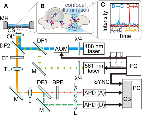
Figure 2. SmFRET using alternative laser excitation (ALEX) is applied to detect the structural distribution and changes of cytosolic proteins in live cells.

(A) Experimental setup. (B) Single-molecule detection of freely diffusing cytosolic molecules in a live cell. (C) Typical fluorescence burst signals. X → Y denotes the fluorescence signal from dye Y with the excitation of dye X, where X and Y are the donor (D) or the acceptor (A), respectively. FG: function generator; λ/4: quarter wave plate; AOM: acousto-optic modulator; M: mirror; DF: dichroic filter; OL: objective lens; MH: metal holder; CS: coverslip; EF: emission filter; TL: tube lens; L: lens; BPF: band-pass filter; APD: avalanche photodiode; CB: counter board; PC: personal computer. Figure reproduced from ref. [25] with permission.
