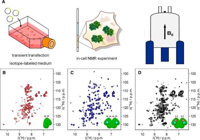
Figure 3. In-cell NMR is applied to detect the conformational changes of cytosolic proteins in live cells.
(A) The vector containing a gene of interest (green arrow) is delivered by transient transfection in labeled medium. Cells expressing the protein of interest (green) are collected and placed in a 3 mm Shigemi NMR tube and applied for in-cell NMR analysis. (B–D) 1H−15N NMR spectra of SOD1 in the apo and reduced state (B), the one-zinc-ion (cyan)-per-monomer bound and dimerized state (C) and the fully mature, copper (salmon)-zinc (cyan)-bound and disulfide-oxidized state (D) in human cells. Figure adapted from ref. [36] with permission. (Further permissions related to this figure should be directed to the ACS. Original figures at: https://pubs.acs.org/doi/10.1021/acs.accounts.8b00147).

