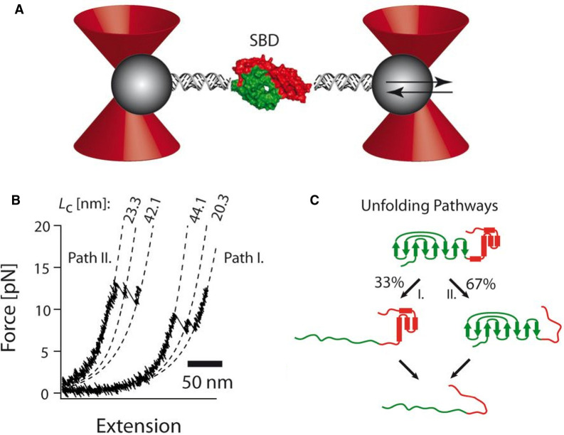
Figure 4. Single-molecule force experiments of the SBD of the Hsp70 chaperone performed with optical tweezers.
(A) Optical tweezers assay. The SBD (green/red surfaces) is tethered to the beads (gray spheres) by two DNA handles, and beads are trapped in highly focused laser beams (red cones). The connection between the DNA and protein is realized by the modification of the two cysteine residues of the protein by the single-stranded DNA-maleimide oligonucleotide complementary to the DNA-handle overhang. One of the beams is reflected by a steerable mirror, which enables pulling and stretching of a single protein. (B) Force-extension curves of a single SBD domain show the order of the individual unfolding events varies. Pathway I corresponds to unfolding of the larger fragment first, followed by the shorter SBD fragment. For pathway II, the order of unfolding events is reversed. (C) Summary of bifurcating unfolding pathways of the SBD. Figure adapted from ref. [56] with permission.

