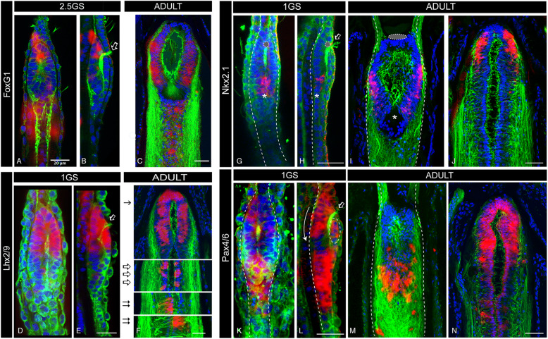
Fig. 3.
Post-metamorphic expansion of domains expressing telencephalic genes in amphioxus. a–c Confocal images of FoxG1 expression in 2.5GS embryos (a dorsal and b lateral view) and adult (c) brains, showing the great expansion of the FoxG1 domain in adults. In embryos, only few cells under the fontal eye are visible (b). c A confocal image of the paraffin section shown in Fig. 2b. d–f Confocal images of Lhx2/9 expression in a 1GS embryo (d dorsal and e lateral view) and an adult (f) brain. In embryos, Lhx2/9 is expressed only in the anterior half of the brain (d, e), while in adults (f) it occupies the entire cerebral vesicle and expands posteriorly into paired clusters of cells, similar to those observed in zebrafish at the midbrain/hindbrain boundary (empty arrows in f) and in the reticulo-spinal system (double arrows in f). f A confocal image of the paraffin section shown in Fig. 2k and l. g–j Confocal images of Nkx2.1 expression 1GS embryos (g, h) and adult (i, j) brains, showing that in early stages of development only a few Nkx2.1+ cells are located in the ventral-caudal side of the brain, close to the infundibular organ (*in g and h). This contrast with the anterior expansion in adults on the ventral side of the brain (i) and a new dorsal-anterior site of Nkx2.1 expression (j), previously undetectable in larval brains. i, j Confocal images of paraffin sections in Additional file 6 Figure S6. k–n Confocal images of Pax4/6 expression in 1GS embryos (k, l) and adult (m, n) brains, showing that the dorsal-anterior domain is greatly expanded in adults (n) while in the larvae is only composed of a few cells (dashed circle in l). m, n Confocal images of paraffin sections in Additional file 5 Figure S5. Empty arrow in b, e, h and l indicates cilia projecting from within the brain through the neuropore, highlighting the brain ventricle that divides the brain into a dorsal and a ventral side in embryos. Colour code: red, gene expression; green, neuropil (acetylated tubulin); blue, nuclear staining (DAPI). All scale bars in amphioxus adult sections are 50μm, while in embryos are 20μm. Abbreviations: 1GS one-gill-slit stage, 2.5GS 2.5 gill-slit stage

