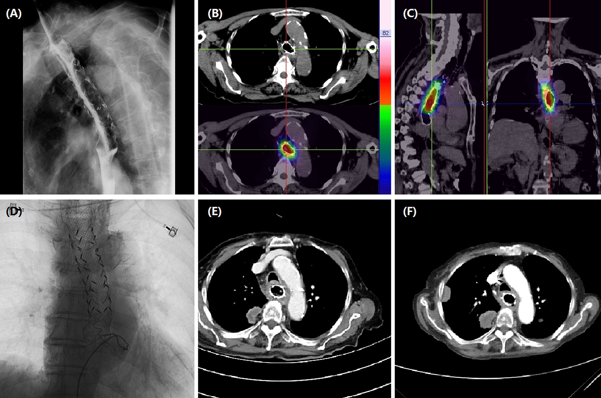
Fig. 4.

Images of an 83-year-old woman with esophageal cancer in upper and middle esophagus. aRadioactive stent was place due to serious stenosis caused by esophageal cancer. bPathological diagnosis of adenocarcinoma. cCT scan shows an obvious thickened esophageal wall with a mass in the right upper lung after 6 months. dThe left gastric artery is super-selectively catheterized and embolized using oxaliplatin loading-CalliSpheres beads. eAfter 9.9 months, CT scan shows a decreased esophageal wall with progressed masses in the right upper lung
