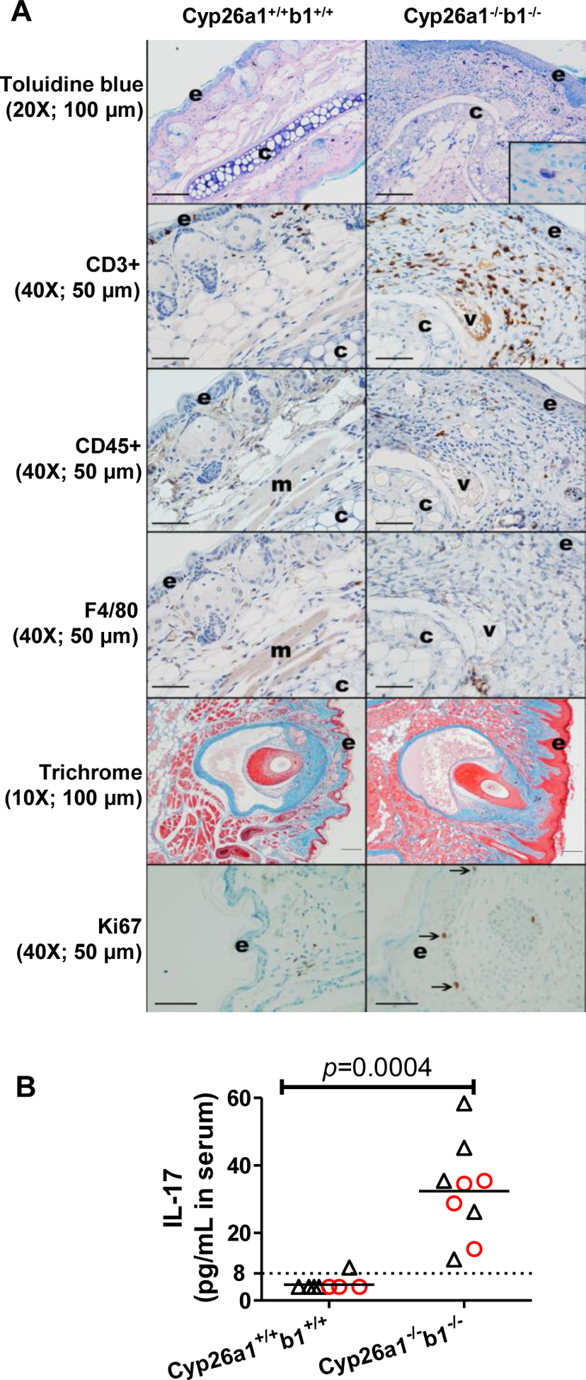
Figure 5.

Representative skin images (A) and serum IL17 concentrations (B). All the data are from the juvenile mice in cohort 1. In panel A, the inset in the top right image shows a mast cell stained with toluidine blue. Arrows in the bottom panels indicate Ki67 positive cells in the stratum basale layer of the epidermis (e); “c” denotes cartilage; “v”, vessel and “m”, skeletal muscle. For CD45, CD3, F4/80 and Ki67 immunohistochemistry, immunoreactive cells are brown; blue = hematoxylin counterstain. Mild nonspecific staining is present in the vessel and skeletal muscle, and some pigment (melanin) is present in the skin. The original image magnification and the length that black scale bars represent are shown inside parentheses. In panel B, each data point represents an individual mouse. Serum IL17 LLOQ (8 pg/mL serum) is indicated by a dashed line. IL17 concentrations were assigned as 4 pg/mL (LLOQ/2) for samples that were below LLOQ. The horizontal lines indicate mean values. Red circles and black triangles represent female and male mice, respectively. The statistical significance was tested by Mann-Whitney test.
