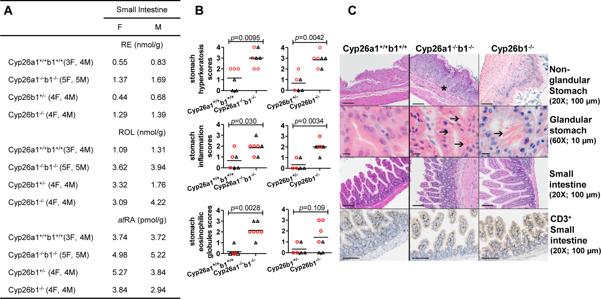
Figure 6.

Concentrations of retinyl palmitate (RE), retinol (ROL) and atRA in the pooled mouse small intestine samples (A). The numbers of female, F, and male, M, mice from the indicated genotypes pooled in each group are shown inside parentheses and values for male and female are reported separately. The histological scores for the stomach hyperkeratosis, inflammation and hyaline droplets (eosinophilic globules) are shown in (B) and each data point represents an individual mouse with red circles and black triangles representing female and male mice, respectively. The horizontal lines indicate mean values and differences between groups were tested by the Mann Whitney test with p-values shown. H&E and anti-CD3 stained images of stomach and small intestine are shown in (C). All the data are from the juvenile mice in cohorts 1 and 2. In panel C, the original image magnification and the length black bars represent are shown inside parentheses. Intracellular hyaline droplets and extracellular eosinophilic crystals in the glandular stomach near the limiting ridge are indicated by arrows and the asterisk indicates inflammation in the nonglandular stomach.
