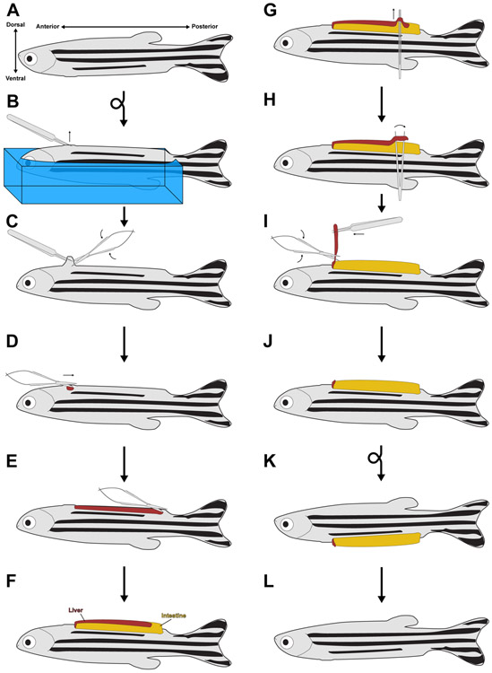
Figure 1. The partial hepatectomy protocol.
(A) Diagram of an adult zebrafish, with the anterior-posterior and dorsal-ventral axes noted. (B) After anesthetization, an animal is embedded in a sponge (shown in blue) so that it is ventral side up. Fine forceps are used to pinch the skin just posterior to the heart. Sponge is not shown in subsequent panels for clarity. (C) Scissors are used to break the skin just posterior to the heart. (D-E) The scissors are used at the now open wound to make a 3-4 mm incision running from anterior to posterior, terminating just before the pelvic fins. (F) Squeezing the sides of the sponge causes the visceral organs to pop out of the larger wound. (G) Fine forceps with both tines touching are slid between the ventral lobe of the liver and the intestine. (H) The forceps are relaxed, causing them to break the portal vein attachments between the live and the intestine. This process is repeated along the length of the ventral lobe. (I-J) The ventral lobe is pulled up from its posterior end, while the scissors are used to sever the ventral lobe from the rest of the liver. (K) The animal is placed into a recovery chamber with system water, where it regains consciousness and rights itself. (L) With time, the viscera are pulled back into the body cavity and the wound heals.

