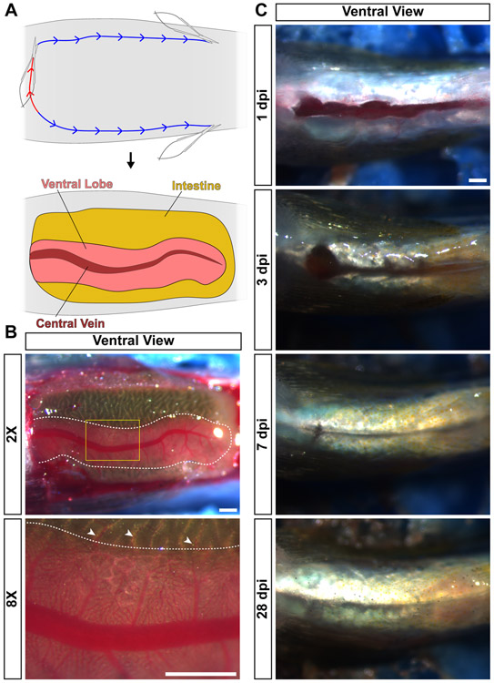
Figure 2. Zebrafish anatomy and recovery from partial hepatectomy.
(A) For analysis of the visceral organs, animals are euthanized and placed ventral side up in a sponge. First, an incision is performed at the anterior-posterior position of the heart (red line). Then, two incisions are generated that run along the anterior-posterior axis to the pelvic fins (blue line). Then, the skin and muscle are peeled back, revealing the visceral organs. Labeled are the intestine, ventral lobe of the liver, and the central vein in that lobe. (B) Live image of a ventral view of a zebrafish prepped for analysis of the visceral organs. The ventral lobe of the liver is outlined with a dotted white line. Yellow box in the 2X image indicates the location of the 8X image. White arrowheads indicate portal vein connections between the intestine and the liver. (C) Ventral view of the wound generated by the partial hepatectomy procedure at the indicated timepoints. Over time, the incision heals completely. Scale bars, 500 μm.

