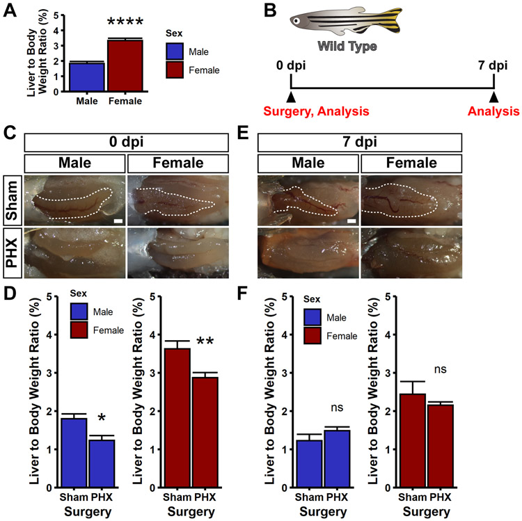
Figure 3. Compensatory regeneration after partial hepatectomy.
(A) Liver to body weight (LBR) ratios were measured for wild type, uninjured zebrafish. Female zebrafish have almost double the LBR compared to male zebrafish. Female animals were compared to male animals using a Wilcoxon rank sum test, ****p<0.0001. (B) Schematic indicating that wild type zebrafish were used for this experiment. Animals were subjected to sham or partial hepatectomy (PHX), and then either fixed at 0 or 7 days post injury (dpi). Fixed animals were imaged and subjected to LBR measurements. (C,E) Shown are representative images of animals subjected to sham or PHX at 0 dpi (C) and 7 dpi (E). Note the complete absence of the ventral lobes in PHX animals. For all images, shown is a ventral view of the visceral organs. Images are bright field images. The ventral lobe of the liver is outlined in white. Scale bars, 500 μm. (D,F) Bar graphs of the liver to body weight ratios for both male and female zebrafish after sham and PHX surgeries at 0 dpi (D) and 7 dpi (F). The height of the bar is the mean value, and the error bars represent SEM. Whereas there is a reduction in LBR following partial hepatectomy at 0 dpi, there is no significant difference between PHX and sham animals at 7 dpi, indicating restoration of LBR. Partial hepatectomy samples were compared to sham controls using a Wilcoxon rank sum test, ns = not significant, *p<0.05, **p<0.01.

