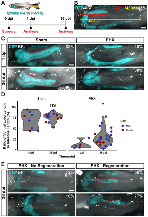
Figure 4. Zebrafish can regenerate their ventral lobes after partial hepatectomy.
(A) Schematic indicating that Tg(fabp10a:CFP-NTR) were used for this experiment. Surgery and analysis by imaging were performed at the indicated timepoints. (B) An example image to demonstrate how animals were analyzed. Animals were quantified by measuring the length of the ventral lobe (in red), the length of the intestine (in yellow) and computing the percentage of the intestine that the ventral lobe occupies (in white). (C) Shown is a representative image of animals subjected to sham or partial hepatectomy (PHX) at 1 and 36 days post injury (dpi). (D) Violin plots of the ratio of ventral lobe length to intestine length for animals subjected to sham or partial hepatectomy at 1 and 36 dpi. Each dot represents the value from a single fish. Values for male fish are in blue, female fish in red. Partial hepatectomy samples were compared to sham controls using a Wilcoxon rank sum test, ns = not significant, *p<0.05. (E) Shown are two examples of partial hepatectomy animals at 36 dpi that did not regenerate, and two examples that did regenerate. For all images, shown is a ventral view of the visceral organs. Images are a merge of a CFP fluorescence image and a bright field image. CFP fluorescence is only present in the liver. The ventral lobe of the liver is outlined in red. The percentage of the intestine that the ventral lobe occupies is in white. Scale bars, 500 μm.

