
Figure 2.
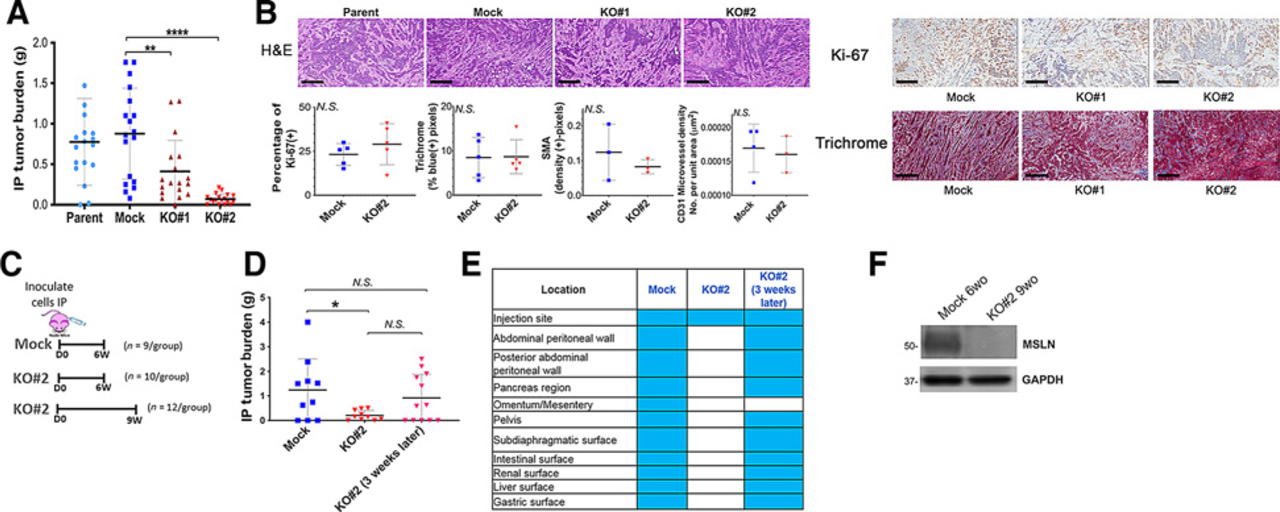
Effect of MSLN KO on intraperitoneal metastasis model. A, Nude mice were inoculated intraperitoneally with 3 × 10⁶ parental KLM1, Mock, KO#1, or KO#2 cells. Mice were euthanized after approximately 6 weeks and all visible tumor in the abdominal cavity was harvested and weighed. Shown are composite results from two experiments (n = 18 for Mock, KO#1 and KO#2; n = 17 for Parent). **, P < 0.01, ****, P < 0.0001 (Mann–Whitney non-parametric test; B), Representative histology stainings of tumors (n = 3–6 mice per group) from (A); scale bars, 300 μm. Samples were analyzed by a consultant veterinary pathologist who found no differences (tumors from n = 3–5 mice obtained from two independent experiments). Quantitation is shown below for staining of Ki-67, SMA (a marker of cancer-associated fibroblasts), collagen on Masson Trichrome and CD31 (a vascular endothelial marker). N.S. = not significant. C, Schema of inoculations for D. D, Experiment in A was repeated but some KO tumors were grown for 3 additional weeks beyond original endpoint (n = 10–12/ group over 3 independent experiments). *, P < 0.05, N.S. = not significant (Mann–Whitney nonparametric test. E, Location of tumor deposits found in animals from D. F, Lysates from harvested KLM1 KO#2 intraperitoneal tumors grown for 9 weeks (9wo) were immunoblotted with anti-MSLN antibody to demonstrate loss of expression of MSLN in vivo. KLM1 Mock intraperitoneal tumors grown for 6 weeks (9wo) were used as positive control for MSLN expression. GAPDH was used as loading control.