
Figure 3.
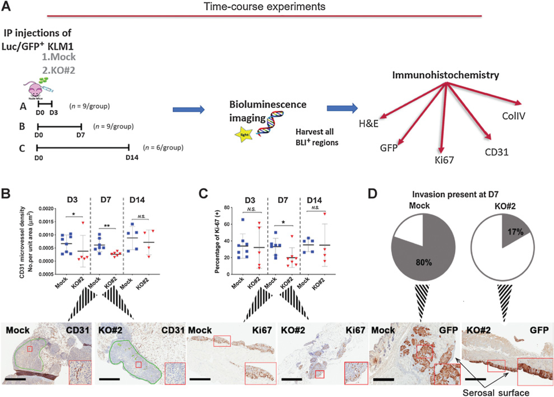
Role of MSLN in establishment of intraperitoneal tumors. A, Schema of time-course experiments. A total of 3 × 10⁶ Luc/GFP cells were inoculated into the mouse intraperitoneal cavity. (n = 4–8/ group over three independent experiments; B–D) Histologic analyses of intraperitoneal tissues containing tumors of (B) microvessel density at 3 (D3), 7 (D7), and 14 (D14) days, green lines show tumor regions selected for pathological analysis (C) proliferation;*, P < 0.05; **, P < 0.01, N.S. = not significant (Mann–Whitney nonparametric test) and (D) invasion of tumor tissue at 7 days after intraperitoneal tumor cell inoculation. 4/5 (80%) Mock and 1/6 (17%) KO#2 tumor tissues showed invasion. Insets (B–D) show magnification of region in red box; scale bars, 600 μm.